Dot Blot Hemorrhages
I find it easiest to find a blood vessel and then follow this vessel back to its origin at the optic disk. Inspect the disk margins and the size of the disk cupping.
Patients With Diabetes Facing Vision Loss At Ever Earlier Ages Acp Internist

Nonproliferative Diabetic Retinopahty Npdr Columbia Ophthalmology
Retinal Hemorrhages Following Fingolimod Treatment For Multiple Sclerosis A Case Report Bmc Ophthalmology Full Text
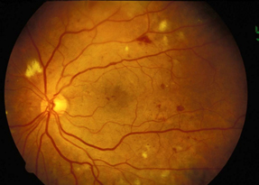
1 mild non-proliferative DR NPDR.
Dot blot hemorrhages. Duration of the diabetes. 27 Referral to a retina specialist is recommended. Sample Question Eye Ear Nose and Throat.
Aside from the obvious flow of blood from a wound or body orifice massive hemorrhage can be detected by other signs such as. Usually blood accumulates in the outer plexiform or inner nuclear layers or more easily seen at peripheral retina where the nerve fiber layer is thin. Click Here for the Correct Answer with an explanation.
It can vary from dot-blot hemorrhages to extensive areas of bleeding that render the underlying sclera invisible. Hemorrhage hemŏ-rij the escape of blood from a ruptured vessel. At least 90 of new cases could be reduced with proper treatment and monitoring of the eyes.
Intraretinal hemorrhages 20 in each of the 4 quadrants definite venous beading in 2 quadrants or intraretinal microvascular abnormalities in 1 quadrant but. Of microaneurysms dot and blot hemorrhages hard exudates or cotton wool spots but less than severe NPDR. DR was graded based on the International Clinical Diabetic Retinopathy Disease Severity Scale as follows.
Managed care noun A popular term for a significant loss of revenue to a providereg a hospital physician office etcresulting from nonreimbusement by third-party carriers for tests or procedures not covered by the insurer or guarantor. Viral infection of the cornea is often caused by the herpes simplex virus which causes a dendritic ulcer with fluorescein stain. Any finding of microaneurysms dot and blot hemorrhages hard exudates or cotton wool spots but less than severe NPDR.
Fluid deposition under the macula or macular edema interferes with the maculas normal function and is a common cause of vision loss in those with DR. They fill the entirety of the retinal layers occupying and displacing the normal retinal architecture therefore forming round uniform hemorrhages. High level of blood total cholesterol and LDL.
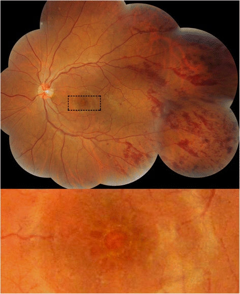
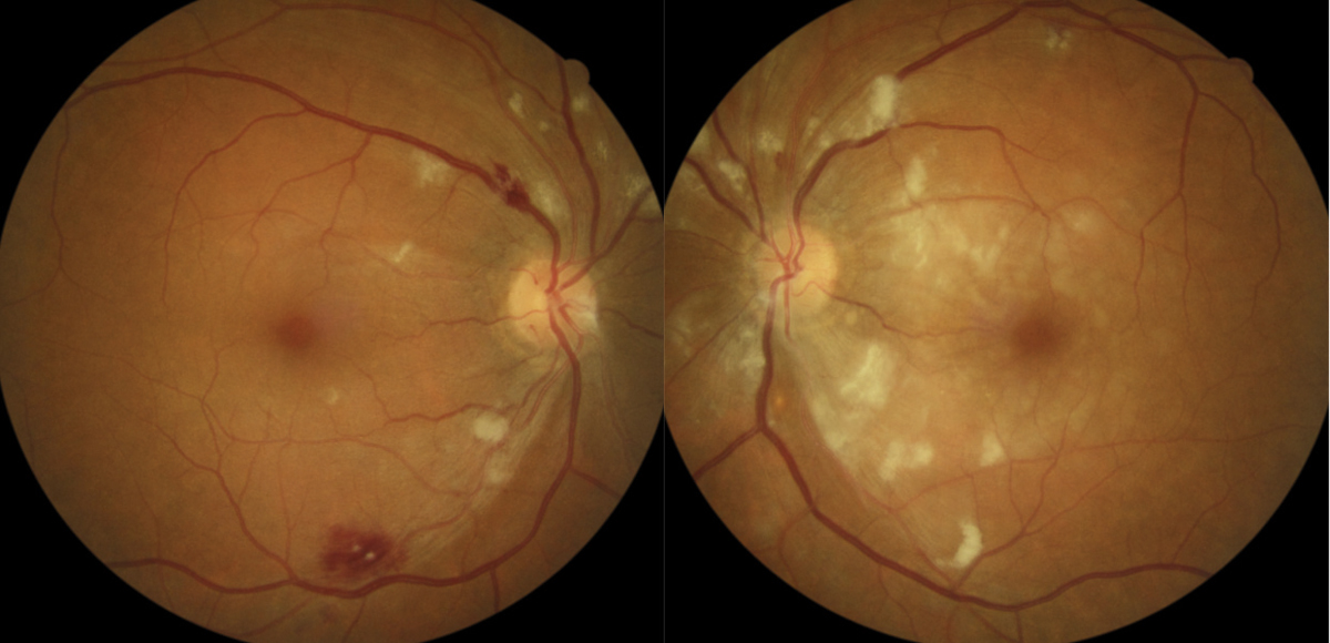
This image of the left retina reveals multiple blot hemorrhages blue arrows of various sizes together with microaneurysms red arrow which can be difficult to distinguish from dot hemorrhages. The fundamental pathologic process involved in capillary occlusion is believed to be the result of an activated leukocyte adhering to and damaging the retinal capillary wall which results in eventual capillary occlusion. Retinal hemorrhages Figure 1-3 develop when necrotic vessels bleed into either the superficial retina nerve fiber layer flame shaped hemorrhage or the inner retina dot blot hemorrhage.
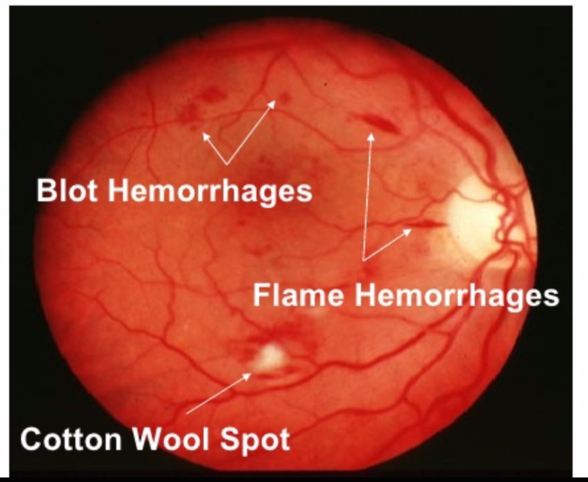
Cotton wool spots Figure 1 3 are caused by ischemia to the nerve fiber layer secondary to fibrinous necrosis and luminal narrowing. Classically there is a wedge-shaped distribution of intraretinal hemorrhage that is more extensive if the occlusion is ischemic compared to a non-ischemic BRVO. Both retinas show scattered intraretinal hemorrhages.
4-2-1 rule -- 4 quadrants of diffuse retinal hemorrhages and microaneurysms 2 or more quadrants of venous beading or 1 or more quadrant of IRMA. Any finding of microaneurysms dot and blot hemorrhages hard exudates or cotton wool spots but less than severe NPDR. Cotton wool spots and hard exudates may be present.
Transposition of great arteries Management Avoid anesthesia-related malignant hyperthermia. 30-34 years of diabetes increase the risk of retinopathy by 65. Infectious keratitis can be caused by bacteria viruses fungi or parasites.
However deeper in the retina since the layers are vertically oriented it results in circumscribed round hemorrhages dot and blot. Diabetic retinopathy affects up to 80 percent of those who have had diabetes for 20 years or more. Intraretinal hemorrhages à 20 in each of four quad-rants definite venous beadingin two quadrants or intraretinal microvascular abnor-.
Bacterial infectious keratitis - improper contact. Retinal hemorrhages are an important ophthalmic diagnostic sign for an underlying systemic vascular disorder. Strict blood glucose blood pressure and cholesterol control.
Race cigarette smoking alcohol. 0 no apparent retinopathy. Laboratory CK High in central core MH patients CK may be high in some asymptomatic carriers G341R In vitro contracture test for malignant hyperthermia Sensitivity.
27 She had heard about Jesus and came up behind him in the crowd and touched his cloak 28 for she said If I but touch his clothes I will be made well 29 Immediately her. The weakened vessels also become leaky causing fluid to seep into the retina. 3 4 The incidence of SCH was reported as 29 in a study with 8726.
Dot and Blot Hemorrhage Hemorrhages lie deeper in the retina. Typical funduscopic examination consists of flame hemorrhages dot and blot hemorrhages cotton wool spots hard exudates retinal edema and dilated tortuous veins Figure 1. Haemorrhage Haematology noun Bleeding which may be pooled or active.
27 The compressive force from the surrounding layers leads to the typical dot or blot shapes Figure 4. It can be either external or internal. 5 Now there was a woman who had been suffering from hemorrhages for twelve years.
Commonly known as dot or blot these are found within the inner nuclear or outer plexiform layers of the retina. Because of their dot-like appearance they are called dot-and-blot hemorrhages. 26 She had endured much under many physicians and had spent all that she had.
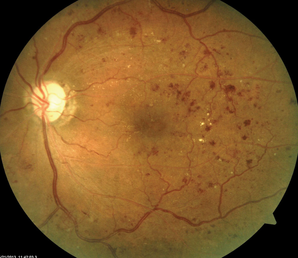
Increased number of microaneurysms and dot-blot hemorrhages. Note the venous beading in two quadrants and the vessel attenuation the dotblot hemorrhages in all four quadrants and the cotton wool spots. Keratitis is a condition in which the cornea becomes inflamed and is often marked by pain impaired eyesight photophobia and red eye.
Blood from an artery is bright red in color and comes in spurts. And she was no better but rather grew worse. You may be able to pick up AV nicking from high blood pressure and retinal hemorrhages in the form of.
If the patient has dot-blot hemorrhages cotton-wool spots venous beading or intraretinal microvascular anomalies IRMAs in the absence of neovascularization classify the DR as nonproliferative. The location size and distribution of the hemorrhages provide clues to the etiology and uncover underlying systemic disorders such as vascular disease hematologic disorders and. DR is classified two ways depending on symptoms.
A 64-year-old overweight man with type II diabetes mellitus is seen for routine examination. 2 Histologically SCH can be defined as hemorrhage between the conjunctiva and episclera and the blood elements are found in the substantia propria of the conjunctiva when a subconjunctival vessel breaks. That from a vein is dark red and comes in a steady flow.
Microaneurysms consequently rupture to form hemorrhages deep in the retina appearing as dots on retinal examination more commonly known as dot and blot hemorrhages. Diabetic retinopathy also known as diabetic eye disease DED is a medical condition in which damage occurs to the retina due to diabetes mellitusIt is a leading cause of blindness in developed countries. Patients with severe NPDR should be monitored using both macular OCT and fluorescein angiography to detect any DME or early neovascularization.
Intraretinal hemorrhages 20 in each of the. They range from the smallest dot and blot hemorrhage to massive sub-hyaloid hemorrhage. Eye Ear Nose and Throat.
Avulsion of retinal vessels and vitreous hemorrhages. Define the severity based on the symptoms. Therefore a superficial bleed will track parallel to the nerve fiber layer resulting in a longitudinal spread becoming flame shaped.
The first step in the pathogenesis. Commonly seen in association with diabetic or hypertensive retinopathy.
My Patient Has Diabetic Retinopathy Now What

Colour Fundus Photographs In Our Case A Dot And Blot Hemorrhages In Download Scientific Diagram
Moran Core Retina Rpe Histopathology

Ophthalmology Notes And Synopses Dr Dot Blot Hemorrhages Photo Credit On Photo Facebook


Diabetic Retinopathy Diabetic Retinopathy 1 Epidemiology And Risk

Optometric Management The Many Faces Of A Retinal Hemorrhage
Busted Barriers Triaging Retinal Hemorrhages
Eye Atlas On Twitter Fundus Findings In Diabetic Retinopathy Blot Hemorrhages Flame Hemorrhages Cotton Wool Spot


Comments
Post a Comment