Ap Pelvis Positioning
The Raz-AP mobile shower commode chair comes standard with 4 of height adjustment an adjustable-tension fabric backrest 5 dual-locking casters flip-up padded armrests and a commode pan. Clarks Positioning in Radiography 12th ed Arnold.

Pelvis Ap View Radiology Reference Article Radiopaedia Org

Ce4rt Radiographic Positioning Of The Hip And Pelvis For X Ray Techs

Diagram Showing Standardised Positioning For Ap Pelvic Radiographs For Download Scientific Diagram
Only Raz offers the Ischial Pelvic Alignment System IPAS which allows the.
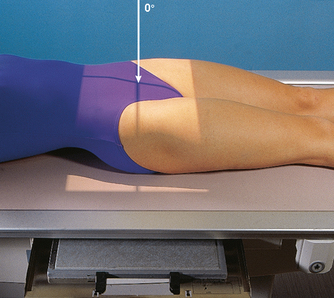
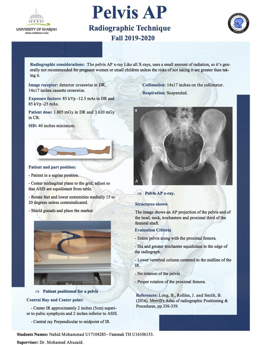
Ap pelvis positioning. Purpose and Structures Shown Clear image of entire pelvis. If a fracture is present your orthopedics colleagues may prefer to manage the injury operatively and any closed manipulation may cause injury to surrounding neurovascular structures. The AP pelvis view is part of a pelvic series examining the iliac crest sacrum proximal femur pubis ischium and the great pelvic ring.
Of both lesser trochanters on the AP pelvis radiograph. Also demonstrates head neck trochanters and proximal one third or one fourth of shaft of femur. 3 Postreduction x-rays should be taken to confirm reduction followed by a computed tomography CT scan in 1- to 3-mm cuts through the pelvis to show concentric.
Positioning Without positioning markers. The 72 views are the AP full spine neutral lateral cervical flexion lateral cervical. Makes appropriate positioning of acetabular component more difficult.
A medial cup will decrease joint reactive forces and decrease force required by abductors to maintain a level pelvis. Initial imaging should include a plain hip with AP pelvis. Indications This view is of considerable importance in the management of severely injured patients prese.
The easy-to-clean polished stainless steel frame is designed for easy side and front access by the user or attendant. 2 In an anterior dislocation the femoral head appears larger than the unaffected hip because the bone is positioned closer to the x-ray source and further away from the film. Examination of the lumbar spine pelvis and hips of men of any age and pre-menopausal females gonad shields should be used.
Indications This projection is utilized. If suspicion for associated fracture subsequent CT is recommended to fully characterize the injury. Measure the distance between the inter-tuberosity line and the line drawn at.
This article discusses radiographic positioning to show the hip and pelvis for the Radiologic Technologist X-Ray Tech. The lumbar spine generally consists of five vertebrae see. A lateral view should be used to confirm this finding.
The lumbar spine anteroposterior or posteroanterior view images the lumbar spine in its anatomical position.
Pelvis And Upper Femora Radiology Key

Radiographic Positioning Of The Femur And Tib Fib Ce4rt
Epos Trade

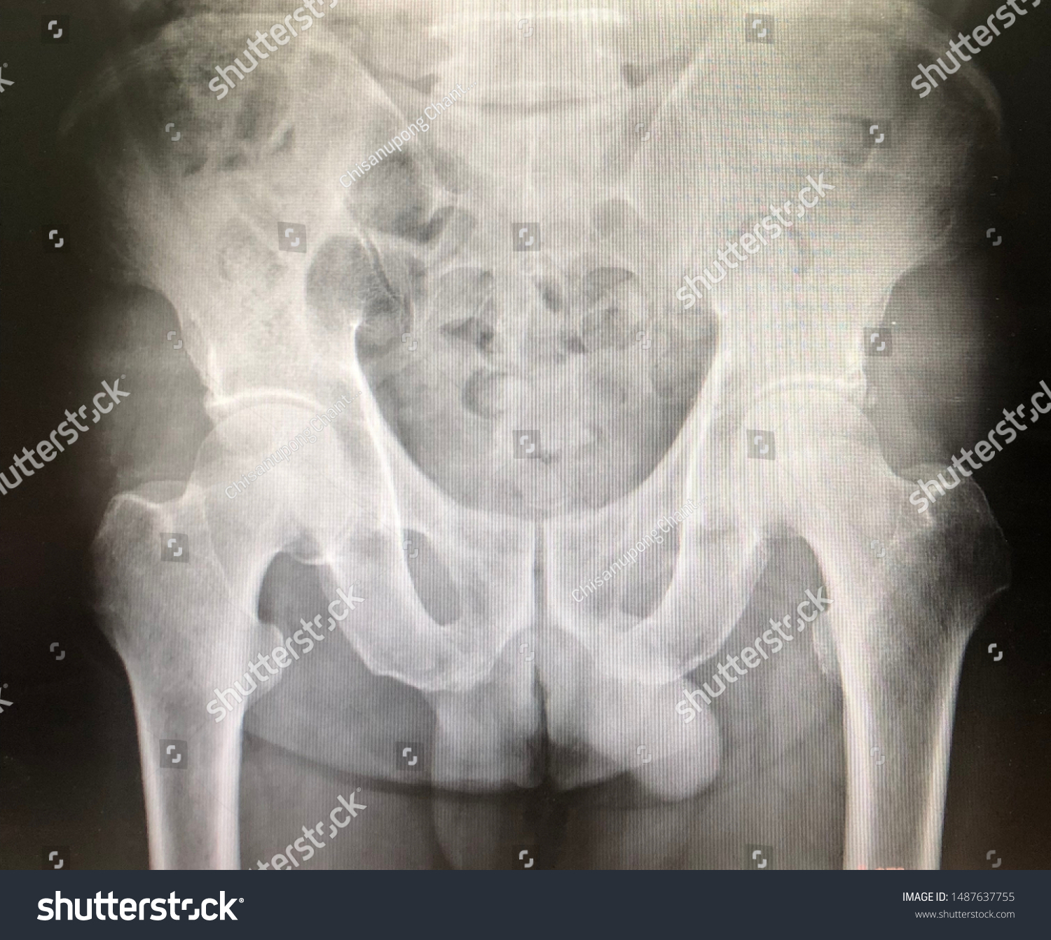
Normal Film Pelvis Ap Position Stock Photo Edit Now 1487637755
Pdf Pelvis Ap Projection

Pelvis And Hips Ppt Download

Proposal For Standardization Of Radiographic Studies On The Hip And Pelvis Sciencedirect

Proper Positioning For The Pelvis And Proximal Femur


Comments
Post a Comment